

Treating blindness caused by burns using limbal stem cells harvested from the undamaged eye of the same patient has now become cheaper, easier and safer. Results of a pilot study of the SLET (simplified technique of limbal transplantation) technique conducted at L.V. Prasad Eye Institute on six patients, and published recently in the British Journal of Ophthalmology provides the proof.
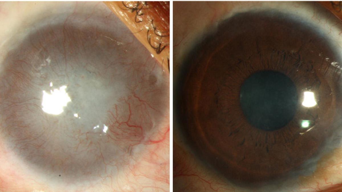
Blindness arises when burns permanently damage the limbal stem cells found in the eye and causes loss in corneal transparency. In such cases, the stem cells are harvested from the healthy eye and transplanted to the damaged eye. There are currently two ways of using limbal stem cells to cure blindness caused by burns.
One is to directly transplant the stem cells to the damaged eye. The other technique — cultivated limbal epithelial transplantation (CLET) — is to remove a smaller portion (2 mm by 2 mm) of the limbus containing the stem cells and increase (expand) the cells in the laboratory and then transplant them to the damaged eye. While both methods are good at restoring vision in the damaged eye, they have their own disadvantages.
In the case of direct transplantation — CLAU (conjunctival limbal autografting), almost 50 per cent of the limbus (6 mm to 8 mm length of the limbus), has to be removed from the healthy eye. Excess removal of stem cells from the healthy eye can permanently damage it.
“At the moment, there is no way of knowing the amount of limbal stem cells found in an [undamaged] eye,” said Dr. Virender S. Sangwan, Head of the Cornea and Anterior Segment Services at L.V. Prasad Eye Institute, Hyderabad. Doctors would come to know of the “deficiency” in the healthy eye in two to three months after the operation. “But how the compromised stem cells will manifest in stressful conditions like an eye infection will be known later,” he explained.
Though the Institute started off by doing direct transplantation (CLAU), it has turned its attention to the safer CLET alternative.
Though this procedure is safer, it is expensive and patients have to visit the hospital twice, one to remove the limbus and the other to transplant the expanded stem cells.
The new technique (SLET) developed recently by Dr. Sangwan and his team at the Institute and Dr. Sheila MacNeil at the University of Sheffield, UK combines the best of both methods.
While only a small portion of the tissue is removed from the healthy eye (as in the case of CLET), the stem cell expansion takes place not in the lab but in the damaged eye itself.
This ensures that the healthy eye is never damaged, the procedure is cheaper and there is less risk of contamination (as the expansion does not take place in a lab). “It would cost only half the earlier procedure (CLET),” he stressed.
If the medium used in the lab provides nutrients for the stem cells, the tear cells do the same job in this case.
The doctors began trying the new technique during the later part of 2009 and performed most of the operations in 2010 and 2011. Altogether 15 cases have been done so far, of which ten patients have already completed six months of observation time post operation.
The procedure
The procedure is quite simple and takes about an hour to perform. In this, the damaged eye is first cleaned and an amniotic membrane is pasted on the cornea using biological glue. The 2 mm by 2 mm limbal tissue harvested from the healthy eye is then cut into eight to nine pieces and placed them on the membrane. Glue is then applied on the cut limbal tissue so that it sticks to the membrane. The eye is then bandaged using soft contact lens.
“The amniotic membrane acts as a scaffold on which the stem cells grow and expand,” Dr. Sangwan explained. “It took the same time [as the CLET technique] for the damaged cornea to be repaired.”
So simple is the procedure that it can be widely adopted by specialists across the country. “With extra training, cornea specialists can perform the operation,” he assured.
